
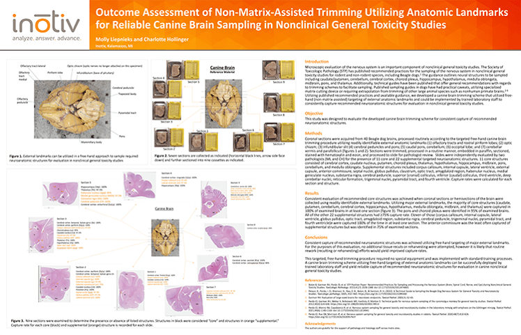
Optimized Free-Hand Canine Brain Trimming Using External Anatomic Landmarks
This study evaluates the efficacy of a non-matrix-assisted, free-hand trimming technique for canine brains in nonclinical general toxicity studies. Using identifiable external landmarks, coronal sections were obtained from 40 Beagle dogs and independently assessed by two pathologists. The method consistently yielded all 11 core neuroanatomic structures, including 100% capture of caudate, putamen, hippocampus, thalamus, and medulla oblongata. This approach is reproducible, does not require specialized equipment, and integrates seamlessly into standard histology workflows—supporting reliable neuropathological assessment in regulatory toxicology settings.
Click here to download the poster.